This patient presented with right upper quadrant abdominal pain. There was RUQ tenderness on exam, but no fever, rebound or Murphy sign. A point-of-care ultrasound was performed to assess for signs of cholecystitis and the following image was obtained. This prompted the operator to ask, “What the heck?”
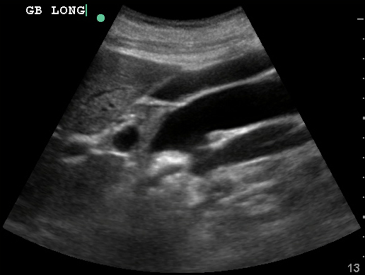
What structures are visible here? How could you differentiate them? More after the break!
As with most ultrasound examinations, fanning through the entire target structure is the best way to appreciate the three-dimensional anatomy and avoid confusion. Here the operator was visualizing the gallbladder as they’d hoped, but the inferior vena cava and aorta photo-bombed their image, causing some confusion.
GB from Sinai EM Ultrasound on Vimeo.
Looking at the video of the gallbladder it is more evident that the aorta and IVC are the culprits, and that we are not dealing with pericholecystic fluid, an aortic dissection, cyst, or some other uncommon pathology. Doppler can be useful in distinguishing vessels in this type of scenario as well. In the image below we can see all the structures labelled, including Gallbladder, IVC, Aorta, and Portal vein. 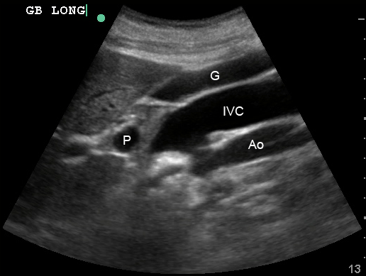
What about the patient, you ask? Normal assessment of gallbladder, normal labs, improved symptoms after an antacid and turkey sandwich. All’s well that ends well.
