Case – 70 year old male mhx of HTN, IDDM presents with 2 hours of exertional chest pain. His ECG in triage is unremarkable for any ischemic changes. He appears uncomfortable and over the next 30 minutes requires escalating doses of nitroglycerin for chest pain. A repeat ECG is unchanged. You decide to echo him to look for a regional wall motion abnormality.
Question – What is the benefit of looking for regional wall motion abnormalities? What is a systematic way to approach this task?
ACS patients often have a non-specific ecg and early negative troponins despite significant coronary occlusion. Regional wall motion abnormalities (e.g. decreased contractility of a specific segment of ventricle) may be the first sign of occult coronary thrombosis. Finding it tunes you into the possibility that the patient may require an early invasive intervention, or at the very least a cath consult.
Cardiologists and trained echocardiographers look at 17 segments of wall motion during an echo. This can be simplified into a 3 part evaluation:
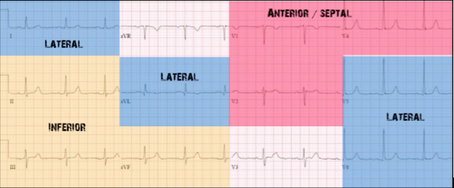
Coronary Artery Territories:
- Blue = lateral wall = L Circumflex (L Cx)
- Yellow = inferior wall = RCA
- Red = anterior wall = LAD


Guidelines:
- Best View – Parasternal Short at level of papillary muscles [3].
- Contractility – can be described as normal, hypokinetic, or akinetic relative to other segments of the ventricle. In a normal ventricular contraction, the segment becomes symmetrically thick as it contracts inward. In areas of dyskinesis, this response is blunted.
- Caveats:
- Distinguishing between old and new regional wall motion abnormalities is very challenging.
- Mechanical and conduction system abnormalities may mimic wall motion abnormalities (e.g. Takotsubos, focal myocarditis, LV Aneurysm)
Anterior Wall Motion Abnormality: (Pathology on L hand side). Source = Regional Wall Motion Asssessment (Teran/Vanyo)
Watch Felipe Teran and Lara Vanyo’s excellent short-talk on this if you’d like to learn more.
Resources:
