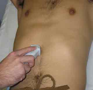
The subxiphoid four chamber view is commonly used in cardiac assessments and the FAST exam and for many is the initial “go-to” view of the heart. Difficulty obtaining this window can frustrate novice and seasoned operators, and there are a few tips which can help optimize the view.
- It’s called SUB-xiphoid for a reason. Don’t jam the probe up against the xiphoid process. Imaging through bone is difficult, the patient will be in pain, and the angle is too steep. Instead, place the probe a few centimeters south of the xiphoid process and work up from there.
- Get a good view of the liver, THEN use that to get a good view of the heart. You may find that starting to the patient’s right of midline gives a better liver window, since the stomach tends to obscure the subxiphoid view as you go further left.
This video illustrates the huge difference that left vs. right can make. It was taken with the probe in a midline subxiphoid position. Starting with the probe angled towards the patient’s left, the entire screen is obscured by gas in the stomach. As the operator changes the angle towards the patient’s right, we see the liver come into view. This yields an excellent window through which the heart can be visualized.
The figure below, taken from the midpoint of the video, illustrates the point a bit more clearly. To the right of the green line (patient left), superficial stomach gas (arrow) obscures everything behind it, creating a terrible view. On the other side of the green line, liver (L) is visualized which creates a good window for viewing the heart behind it.


