74 yo F with hx of CAD, afib, HLD, severe LV dysfunction is brought in by EMS as a notification for a STEMI. A 12-lead EKG in your ED shows the same as the EMS EKG. Should you activate the CATH lab?
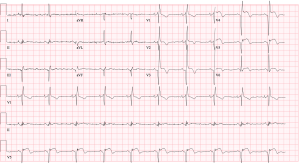
Answer: Check an old EKG.
An old EKG 3 months before showed the exact same EKG morphology. A Cath report from that prior hospitalization showed stable disease with an LV aneurysm.
Why do LV aneurysms present with persistent ST-Elevation? There is some thought that it represents incomplete reperfusion and subsequent scar tissue formation. These persistent ST-elevations are usually seen in the precordial leads and are accompanied by deep q or qs wave complexes and t-wave inversions:
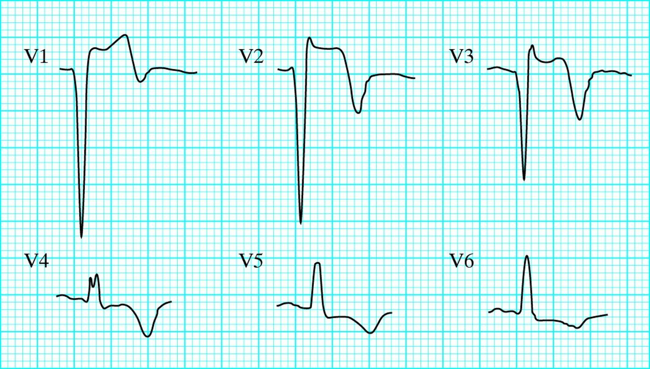
In a patient with STE on EKG without reciprocal changes, appears well, and has a previous last EKG of similar morphology, consider the diagnosis of LV aneurysm. A bedside ultrasound will demonstrate a discrete dyskinetic area of the LV.
Example of an apical 4 chamber view cardiac US demonstrating an apical LV aneurysm (similar to this patient) by Dr. Bandano at http://3decho360.com/cc01/

Sources:
https://lifeinthefastlane.com/ecg-library/left-ventricular-aneursym/
