A couple very brief tips from Sinai Ultrasound QA today.
Views: For each indication a specified set of views are required. Please be sure to obtain all of the required views if you are recording the study – whether for clinical or educational documentation.
Full ultrasound tutorials covering these studies can be found at sinaiem.us/tutorials.
But here is a quick review of the views required for the major ultrasound indications:
- Cardiac: (2 images): subxiphoid, parasternal long axis, parasternal short axis or apical.
- FAST: (4 images): hepatorenal recess (Morison’s Pouch), perisplenic (Left Upper Quadrant), subxyphoid or parasternal long axis cardiac view, bladder.
- Gallbladder: (2 images): transverse, sagittal
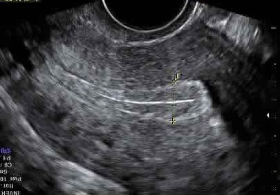
- OB: (2 images): transverse, sagittal views of uterus.
- Aorta: (5 images): proximial, mid and distal aorta (transverse), bifurcation of the iliac arteries (transverse), and longitudinal shot of mid-aorta.

Be sure to center the organ of interest on the screen so that the area deep
to the structure is imaged. This is important to document the absence or
presence of pericardial effusion deep to the heart in a cardiac study, free
fluid deep to the bladder on a FAST exam, or free fluid in cul-de-sac on OB
study.
