Introduction:
The first step in evaluation for pneumothorax (after the physical examination) has traditionally been the use of chest radiography. Both the chest x-ray and physical exam, however, have significant downsides. Fortunately for those patients who can’t be sent immediately to CT scan, ultrasound has been shown to be far more sensitive in the detection of pneumothorax.
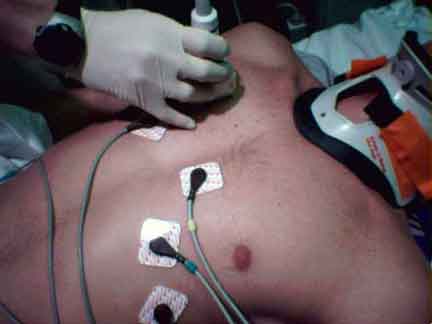
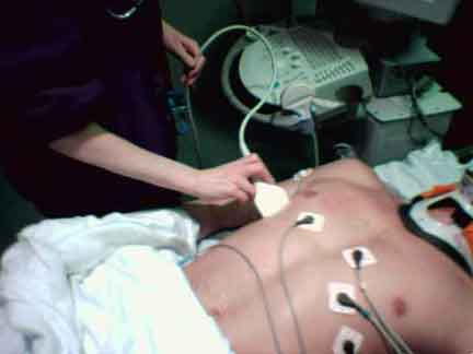
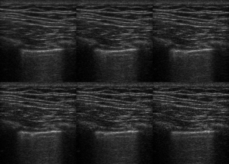
To evaluate for pneumothorax with ultrasound we look for the absence of a normal lung and pleural interface. Using a linear transducer, first the pleural line is identified. Signs of normal lung and pleura are lung sliding and comet tails. Thus the absence of lung sliding or comet tails makes the ultrasound diagnosis of pneumothorax. Since this evaluation is particularly dynamic a clip can be saved, or M-mode may be used to record evidence of normal lung or pneumothorax. We will describe these techniques below.
Focused Questions:
1. Is there absence of lung sliding/comet tails on the affected lung field?
Required Views:
1. Second to fourth intercostal space, bilateral lung fields
2. Additional views should be obtained down anterior chest wall.
Probe Selection:
 |
Linear array transducer(Small parts/high frequency probe) |
Probe Positioning:
Normal Anatomy:
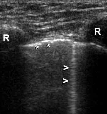 |
Rib shadows (R) are visible as bright reflectors with distal shadow.
The Pleura (* *) is a bright echogenic line beneath the ribs.Comet Tail artifacts (> arrows) arise from normal pleura reflecting sound waves. |
Using Video:
Using M-Mode:
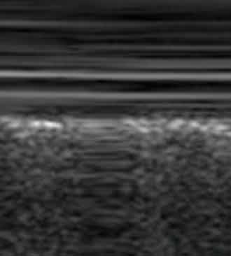
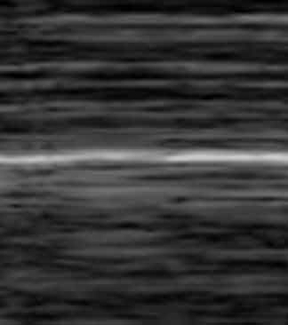
M-mode allows documentation of lung sliding with a single image. Superficial soft tissue/muscle does not move much during respirations, and will yield flat lines on M-mode (top half of images below). Normal lung sliding creates a grainy M-mode appearance (left image, bottom). This is referred to as “waves on a beach,” where the smooth soft tissue lines (top half) meet the rough lung lines (bottom half) at the bright white pleural line (center). With pneumothorax, lung sliding is absent- flat lines will be seen above and below the pleura.
| Normal Lung: Smooth lines above pleural line, grainy below | Pneumothorax: Smooth lines above and below pleural line |
 |
 |
