Introduction:
During the first trimester of pregnancy, evaluation for complaints of lower abdominal pain or vaginal bleeding should include an assessment for ectopic pregnancy. Bedside pelvic ultrasound to detect an intrauterine pregnancy (IUP) can speed this evaluation. When an IUP is visualized, ectopic pregnancy can be ruled out (unless there is suspicion for heterotopic pregnancy). Ectopic pregnancy cannot be excluded in a pregnant patient without a definitive IUP on ultrasound.
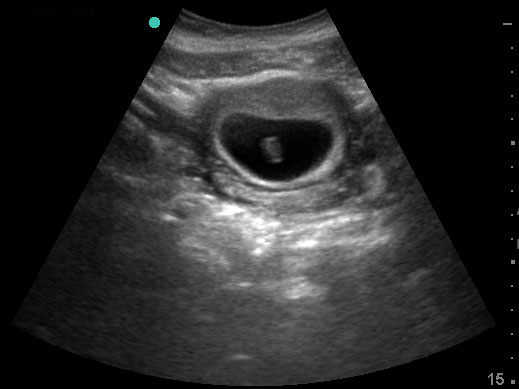
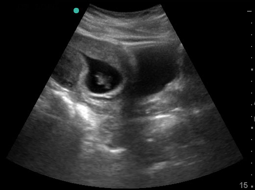
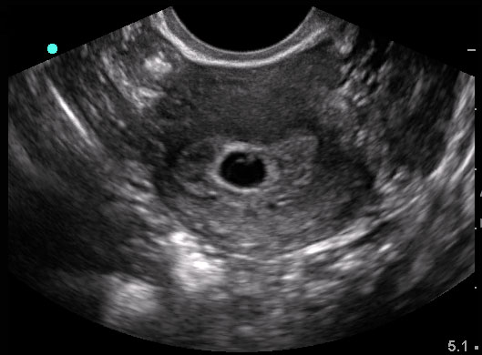
Although a double decidual sign and gestational sac have been described in the obstetrics and radiology literature as the earliest signs of a true IUP, the double decidual sign is inconstant and many false positives exist. It is difficult to appreciate the difference between a true gestational sac and a pseudogestational sac (a collection of fluid or blood in the uterus which mimics early pregnancy but is NOT an IUP). Thus, most emergency physicians will not define a true IUP until a yolk sac is seen.
Focused Question:
- Is there an intrauterine pregnancy?
Video Overview:
Required Views:
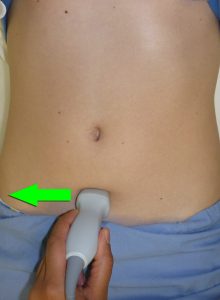
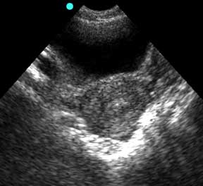
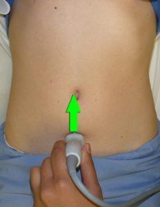
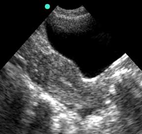
Always start with a transabdominal approach. This gives the best overall, “big picture” view of pelvic anatomy and is especially useful when the gestational age is unclear at the start of the evaluation. Also, ensure that the uterus has been thoroughly visualized in transverse and saggital planes prior to any evaluation of the ovaries.
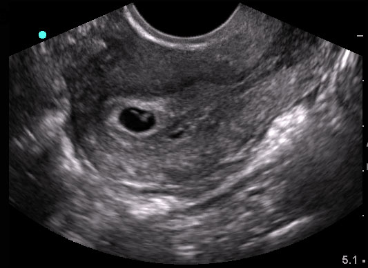
When a suspected IUP is visualized, it is critical to double-check that it actually resides within the uterus. Myometrium should surround the entire gestational sac. The thickness of the myometrium surrounding the sac, also known as the myometrial mantle, should be at least 8mm thick. The uterus should be demonstrated from cervix to fundus in two planes.
1. Transverse view of uterus (Transabdominal approach)
2. Longitudinal view of uterus (transabdominal approach)
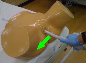
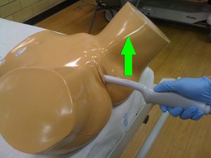
3. Transverse or coronal view of uterus (Transvaginal approach)
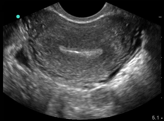
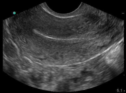
4. Longitudinal view of uterus (Transvaginal approach)
