Introduction:
In the setting of suspected renal colic, bedside ultrasound can assist in making the diagnosis by assessing for hydronephrosis. The goals of bedside ultrasound are to look for the presence and extent of hydronephrosis, assess urine volume, and perhaps look for ureteral jets as well. If abdominal aneurysm is in the differential diagnosis, it is important that AAA be assessed as well. Some aneurysms or other masses can cause hydronephrosis, so it does not exclude the diagnosis of AAA.
Focused Questions:
- Is there hydronephrosis?
- What is the bladder volume?
Video Overview:
Required Views:
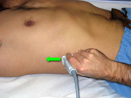
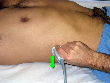
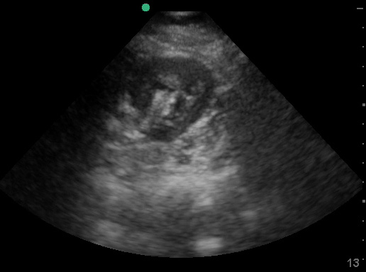
1. Right Kidney (Longitudinal)
| Probe position | Image |
 |
 |
| Notes |
|
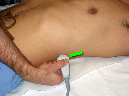
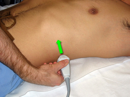
2. Right Kidney (Transverse)
| Probe position | Image |
 |
 |
| Notes |
|
3. Left Kidney (Longitudinal)
| Probe position | Image |
 |
 |
| Notes |
|
4. Left Kidney (Transverse)
| Probe position | Image |
 |
 |
| Notes |
|
Abnormal Image Gallery:
5. Bladder (Transverse)
| Probe position | Image |
 |
 |
| Notes |
|
6. Bladder (Longitudinal)
| Probe position | Image |
 |
 |
| Notes |
|
Estimating bladder volume
Many machines contain built-in calculators for bladder volume estimation based on measurements. One simple and validated equation is simply:
length x width x height x 0.75
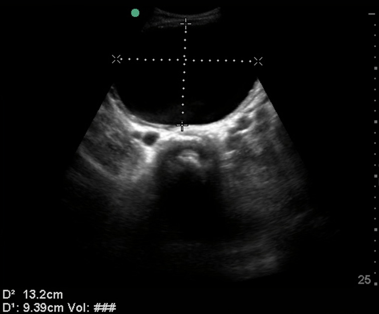
Stated another way, 75% of the cubic volume of the bladder. To measure all three axis of the bladder you will need a transverse view in largest dimension (for length [anterior-posterior] and width [left-right]) and a sagittal measurement (for length and height [cranial-caudal]).
This estimation can be helpful in determining post-void residual noninvasively, in the evaluation of anuria, renal failure, hydronephrosis, Foley catheter placement, and other renal and bladder anomalies. In infants, measuring the bladder volume prior to catheterization reduced the ‘dry tap’ rate significantly.
