Introduction:

Bedside sonography can greatly enhance the evaluation of patients with suspected cholecystitis. Findings of gallstones or a sonographic Murphy sign (maximal tenderness when pressure is applied over the visualized gallbladder) increase the likelihood of cholecystitis. Other sonographic findings which can sometimes be seen include gallbladder wall thickening (greater than 3 to 4mm is a commonly used cutoff), common bile duct dilatation (greater than 5 to 6mm), or the presence of pericholecystic fluid.

Focused Questions:

  1. Are there gallstones?
  2. Is there a sonographic Murphy sign?
  3. Are there other signs of acute cholecystitis?
    • Gallbladder wall > 3 mm
    • Common bile duct > 6 mm
    • Pericholecystic fluid

Video Overview:

Required Views:

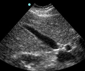
1. Longitudinal View of Gallbladder

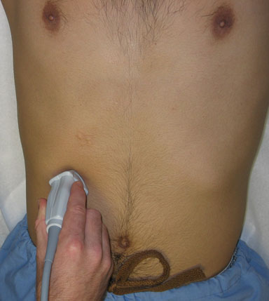
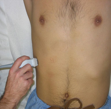
Probe position options: Image
Gallbladder- subcostal approachGallbladder- intercostal approach Gallbladder- long axis
Notes
  • Visualize the gallbladder, portal vein, and common bile duct (if visible)
  • The probe orientation (transverse vs. longitudinal) depends on the gallbladder orientation
  • In most patients, oblique probe orientations will be needed to visualize the “long” and “transverse” views
Abnormal Studies


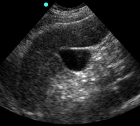
2. Transverse view of gallbladder

Probe position options: Image
Gallbladder- subcostal approachGallbladder- intercostal approach Gallbladder- short axis
Notes
  • The probe orientation (transverse vs. longitudinal) depends on the gallbladder orientation
  • In most patients, oblique probe orientations will be needed to visualize the “long” and “transverse” views
Abnormal Studies
April 2024
M T W T F S S
1234567
891011121314
15161718192021
22232425262728
2930  

Archives