54M with h/o HTN, DM, Tobacco and Focal Segmental Glomerulosclerosis presents with neck mass. He looks dyspneic and uncomfortable at triage and has an obvious mass above his left clavicle to the degree that his head is tilted a bit to the right. Concerned, the triage RN defers the EKG and A-Side attending consult and rolls the patient into your formerly mellow cardiac room shift. Although overall he looks gaunt, his face is swollen and dark colored (“facial plethora”). You go through your IV/O2/Monitor/ABCDEFGHIJKLMNOPQRST and then grab the ultrasound machine.
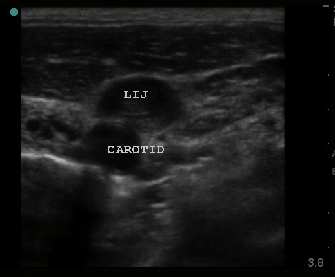
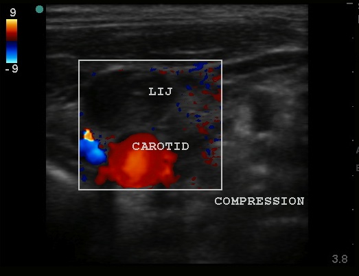
After turning on the machine, you obtain the following images of the left neck.


What do you think the patient has and whatcha going to do about it?
Your suspicions from the patient’s history and physical are confirmed by the ultrasound findings of thrombus in the IJ. This patient very likely has Superior Vena Cava syndrome. The best study would probably be a CTA but with his history of renal disease and your ultrasound findings, you decide not to give him a contrast dye load for further confirmation. Your patient is admitted to the hospital. The next day he undergoes MRA/MRV and cervical node biopsy which confirms neoplasm and a course of radiation and chemotherapy is prescribed.
