Bedside sono for DVT: Ready for primetime?
You got a patient with an enlarged, red, angry leg. It screams sono me for DVT! It is midnight and radiology tells you it cannot be done until the morning. Can YOU sono the patient?
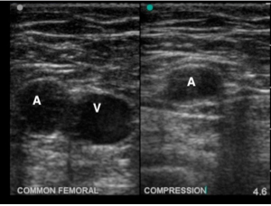
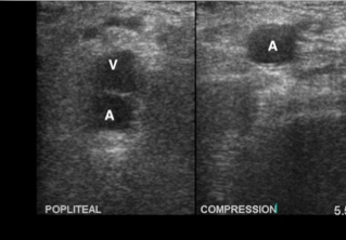
Technically yes…we have an ultrasound and bedside sono’s for DVTs are easy right? 2-point compression-meaning compression of the common femoral and popliteal veins alone is all we need. According to Jang et al 2010 2-point compression had a S&S of 100%/99% respectively when compared with radiology department studies. That was amongst ED physicians at various levels of training who had received a 10-min tutorial on bedside sonos.
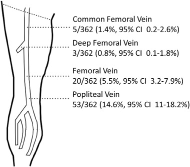
More recently the literature such as Adhikari et al 2015 shows we may miss approx 6% of DVTs w/ the 2-point approach. Primarily in the femoral vein and deep femoral vein.
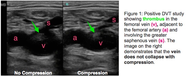
As Mike, Mike, and Matt point out on their Ultrasound Podcast non-ambulatory patients are more likely to have occult DVTs (segmental or incompletely occlusive) and in areas not captured by 2 point compression. Also in general it is very common to have DVTs at venous confluences such as the saphenofemoral branch which may be missed in 2-point compression.
According to a conversation with Adhikari he instructs evaluation at the following areas: (1) common femoral vein (2) saphenofemoral junction (3) deep femoral vein (4) popliteal and its branching points.
See Ultrasound Podcast for how to perform Whole Leg US
RECAP:
- Make sure you’re checking for DVTs at branch points such as saphenofemoral branch point
- In non-ambulatory patients be weary of DVTs in segmental areas or areas not captured on 2-point bedside sono
- Of course your pretest probability will ultimately factor into your management for each patient
Sources:
- Crisp JG, Lovato LM, Jang TB. Compression ultrasonography of the lower extremity with portable vascular ultrasonography can accurately detect deep venous thrombosis in the emergency department. Ann Emerg Med. 2010 Dec;56(6):601-10.
- Adhikari, S., Zeger, W., Thom, C., & Fields, J. M. (2015). Isolated deep venous thrombosis: implications for 2-point compression ultrasonography of the lower extremity. Annals of emergency medicine, 66(3), 262-266.
- Lin, M. PV Card: Focused Deep Vein Thrombosis (DVT) Ultrasound. https://www.aliem.com/2015/pv-card-focused-deep-vein-thrombosis-dvt-ultrasound/
- Dawson, M., Mallin, M., & Stone, M. DVT Ultrasound Controversy! @bedsidesono discusses why 2 point compression not good enough #foamed
http://www.ultrasoundpodcast.com/2015/04/dvt-ultrasound-controversy-bedsidesono-discusses-why-2-point-compression-not-good-enough-foamed/?utm_source=feedburner&utm_medium=feed&utm_campaign=Feed%3A+EmergencyUltrasoundPodcastPodcast+%28Ultrasound+Podcast%29 - Dawson, M., Mallin, M., & Stone, M. DVT Ultrasound demonstration with @bedsidesono. The whole upper leg approach. #FOAMED http://www.ultrasoundpodcast.com/2015/05/dvt-ultrasound-demonstration-with-bedsidesono-the-whole-upper-leg-approach-foamed/?utm_source=feedburner&utm_medium=feed&utm_campaign=Feed%3A+EmergencyUltrasoundPodcastPodcast+%28Ultrasound+Podcast%29
