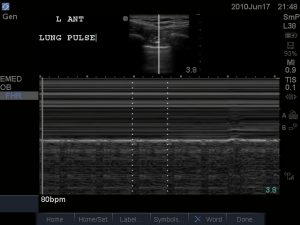
The “lung pulse†is an ultrasound sign first described by Dr Daniel Lichtenstein in 2003. Essentially, it is the detection of the subtle cardiac pulsation at the periphery of the lung (parietal pleura to be exact) on the M mode.
In a normally ventilating lung, this subtle transmission is present but NOT seen, as it is masked or “erased†by the air artifact generated by lung sliding. In a non-ventilated lung, however, lung sliding is abolished. Here, the pleura is perfectly still and this allows the underlying cardiac pulsation to be detected.
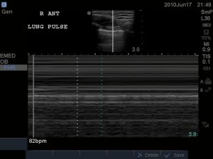
Since the heart is on the left side, lung pulse is more prominent on the left side than right (both images taken from a healthy volunteer while holding his breath).
Thus, for the lung pulse to be seen on M mode, the following two conditions must be present:
1. Absent ventilation (i.e. no lung sliding)
2. Apposition of visceral pleural and parietal pleural (i.e. no pneumothorax)
The clinical utility of the presence of a lung pulse is:
- Diagnosis of non-ventilated lung (sensitivity of 93% and specificity of 100% in patients without previous respiratory disorders)
- Exclusion of pneumothorax
Ref:
- Lichtenstein et al (2003).The “lung pulseâ€:an early ultrasound sign of complete atelectasis.  Intensive Care Med. 2003 Dec;29(12):2187-92. http://www.ncbi.nlm.nih.gov/sites/pubmed/14557855
- Lichtenstein. Pneumothorax and introduction to ultrasound signs in the lung. In: General ultrasound in the critically ill. Springer, pp110-111
