Tl;dr: (1) Never rock the pelvis. Firmly squeeze and hold. (2) Consider quickly assessing for rectal or vaginal bleeding prior to binder application as this would suggest an open fx into the vag / rectal vault. It will be difficult to complete the exam once the binder is on. (3) The binder is applied over the greater trochanters, NOT the iliac crest. Incorrect positioning of the binder can make fractures worse and provides suboptimal stabilization of the pelvis.
You’re walking by the trauma bay when you hear a member of the trauma team shout out that the pelvis is unstable and you see a blood pressure of 80/49 on the monitor. You realize that things are about to get hairy, and interesting, so you decide to grab the pelvic binder for the team…
We are taught to reach for the pelvic binder as one of the first moves in a hemodynamically unstable pt with pelvic trauma. The goal is to decrease pelvic volume in an effort to create a tamponade effect. The binder has secondary benefits of stabilizing the fracture and providing patient comfort.
The examiner should keep a few things in mind prior to applying the binder. First, never ‘rock’ the pelvis on examination as it might shear additional blood vessels and subsequently increase bleeding. Instead, the examiner should gently squeeze and firmly hold the pelvis in place upon recognizing instability. Letting the pelvis fall back into resting position may also cause further blood vessel trauma.
The team may also consider quickly assessing the vaginal cavity and rectal vault for blood. Roughly 5% of pelvic fractures are open with the presence of rectal or vaginal tears, and will require broad spectrum antibiotics and close monitoring for sepsis. Regardless, hemodynamic stabilization takes priority and the complete examination can conceivably be deferred to the OR setting.
A common mistake is to place the binder over the iliac crests, which may exacerbate fractures and obstruct access to the abd for laparotomy. Instead place the binder over the greater trochanters for optimal mechanical stabilization. After application, the lower extremities may be taped in internal rotation, which decreases the anatomic space in the pelvis and augments the impact of the binder.
Finally, do not apply excessive force in tightening the binder. Certain fracture patterns (particularly ones with a lateral compression pattern through the pubic rami) will get worse with the binder. Get ortho involved quickly, they may even request removal of the binder once imaging is obtained.
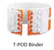
Side note: we have the T-POD binder at Elmhurst, see below. There is no evidence that the device is better than a regular old sheet. In fact, some experts have criticized the T-POD given its size which often limits access to either laparotomy or angiography (and in some cases both). If surgery is available, ask if they prefer a sheet to the T-POD.
Sources:
Brohi, K. The Ideal Pelvic Binder. Trauma.org. http://www.trauma.org/index.php/main/article/657/. Accessed Aug 2018.
Fiechti JF, Gibbs, MA. An Evidence-Based Approach To Managing Injuries Of The Pelvis And Hip In The Emergency Department. EBMedicine.net. December 2010 Volume 12, Number 12.
