Question: What are you imaging?
Answer: What ARE you imaging?
Always keep an eye on the depth you are scanning. Doing so can keep you from mistaking a structure of a different size from the structure you are expecting to find.
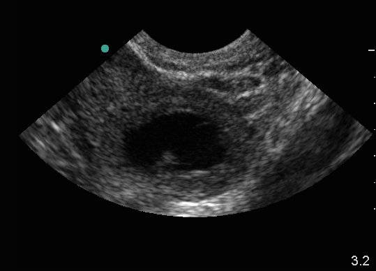
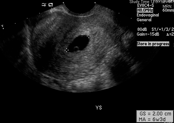
The first ultrasound (Fig 1) was performed at the point-of-care in the ED, and the next image (Fig 2) was a formal ultrasound performed on the same patient in radiology. They show structures of similar appearance, but there are a few clues that these are not the same structures.
The first clue is the size of the probe footprint at the top of the screen. Although both are transvaginal ultrasounds with tightly curved transducer footprints, the ED scan has a much larger footprint indicating that it is a “close up.” The second clue is the actual depth itself which is reported on the bottom right of the image (Fig 1). The full depth of field is only 3.2 inches, which is very shallow for a full view of the uterus. In contrast, the image in Fig 2 is at least 6 cm deep (each dot is one cm).

The very shallow ED ultrasound identifies a cystic structure containing an internal echo. The differential diagnosis of this image includes ovarian cyst, ectopic pregnancy or intrauterine pregnancy (IUP). Note that the myometrial mantle in the Fig 1. would be quite thin, even if we were to call that structure the uterus (which it is NOT). A myometrial mantle less than 8 mm is unlikely to be seen with IUP, and should prompt evaluation for ectopic or interstitial pregnancy. Thus, measuring the myometrial mantle for each of these first trimester bleed/pain patients is an excellent second check that your “IUP” is indeed intrauterine.
The radiology image is at an appropriate depth and shows a different structure, the true IUP in the true uterus. Figure 1 represents the corpus luteum with an internal echo.
Pelvic ultrasound summary tips:
- Start with a large depth of field, and “close in” on your target anatomy once you’ve identified it well
- Scan through the entire uterus in long and transverse/coronal planes fully before looking to the adnexa or focusing in on a possible IUP
- Check behind the uterus for free fluid in the Pouch of Douglas
- If you see an IUP, measure the myometrial mantle as a check against interstitial ectopic pregnancy and to verify that you are truly within the uterus
- Pregnant patients with lower abdominal pain or vaginal bleeding are at risk for ectopic pregnancy. If no IUP is visualized on ultrasound this diagnosis has not been ruled out, regardless of the quantitative hCG level.
