Anatomy
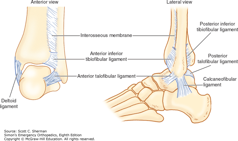
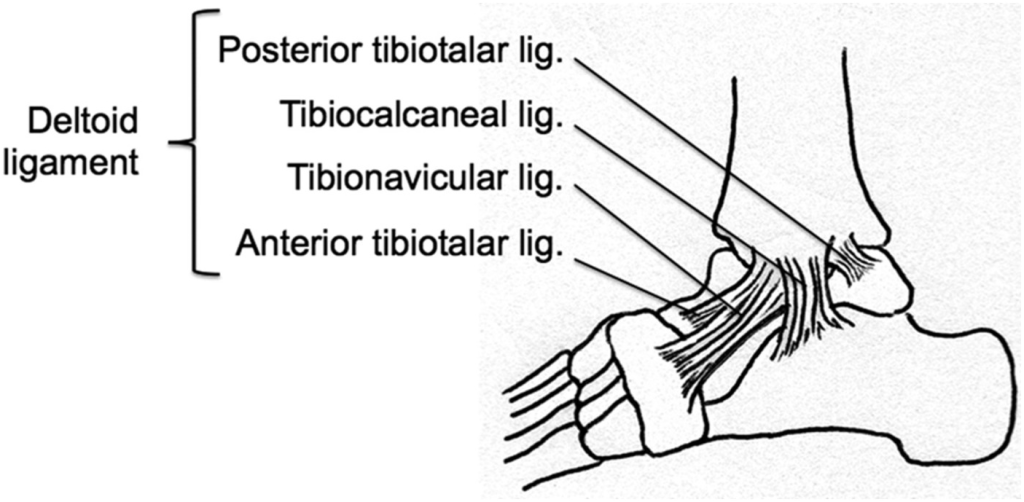
First, we’ll refresh our bony anatomy to contextualize the radiographic images. The ankle is composed of the distal tibia and fibula and the superior talus, which is stabilized by several ligaments and the fibrous syndesmosis.
The standard ankle radiographic series consists of the AP, mortise and lateral views.

The AP View
- Evaluates the ankle mortise, lateral process of the talus and soft tissue edema adjacent to the malleoli
- Image is obtained with the foot in dorsiflexion
- The adequate AP view should have:
- Distal tibiofibular overlap (normal is >6 mm)
- The lateral and medial malleoli in view
- Tibiotalar joint space open, but the mortise is not completely visualized
- If the mortise if completely visualized then it is an AP mortise view
The Mortise View
- Evaluates the ankle mortise, which is made up by the articulation of the tibia and two malleoli with the talar dome
- Image is obtained with the leg in internal rotation at 15-20 degrees with the foot in slight dorsiflexion
- Normal findings:
- Joint space width is uniform and measures 4 mm throughout
- Assessing the talus:
- Surface of the talar dome is smooth without irregularities
- Medial and lateral tubercles of the talus are intact
- Assessing the tibiofibular interosseous membrane
- Tibia and fibula overlap to some degree
- Separation here suggests a torn interosseous membrane
- The space between the distal fibula and tibia at 1-cm proximal to the tibial articular surface should measure ≤ 6mm
- Measurements > 6mm suggest a torn interosseous membrane
- Tibia and fibula overlap to some degree

The Lateral View
- Evaluates for fractures, dislocations, joint effusions and calcaneal fractures
- Image is obtained with the patient in a lateral decubitus position and foot in dorsiflexion
- Normal findings:
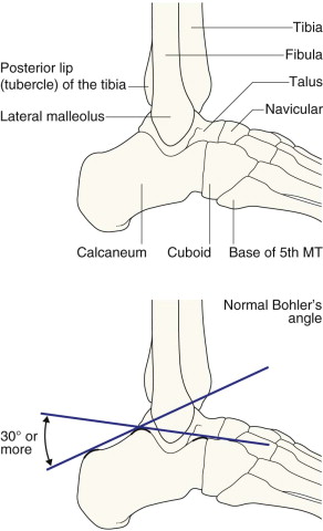
- The lateral malleolus is superimposed on the medial malleolus and extends more inferiorly than the medial malleolus
- The posterior tubercle (posterior malleolus) of the tibia is visualized
- The calcaneum is demonstrated and used to measure Bohler’s angle (normal >30°)
- A reduced Bohler’s angle suggests a calcaneal fracture
- Base of the 5th metatarsal is included
References
Davenport M, Galasso MM. Ankle. In: Sherman SC. eds. Simon’s Emergency Orthopedics, 8e. McGraw Hill; 2019. Accessed January 04, 2023. https://accessemergencymedicine-mhmedical-com.eresources.mssm.edu/content.aspx?bookid=2455§ionid=193455124
Feger J, Knipe H, El-Feky M, Tibiofibular overlap. Reference article, Radiopaedia.org (Accessed on 04 Jan 2023) https://doi.org/10.53347/rID-77885
Gorton S, Jones J, Bell D, et al. Ankle (AP view). Reference article, Radiopaedia.org (Accessed on 04 Jan 2023) https://doi.org/10.53347/rID-40728
Gorton S, Jones J, Bell D, et al. Ankle (mortise view). Reference article, Radiopaedia.org (Accessed on 04 Jan 2023) https://doi.org/10.53347/rID-40730
Gorton S, Bell D, Er A, et al. Ankle (lateral view). Reference article, Radiopaedia.org (Accessed on 04 Jan 2023) https://doi.org/10.53347/rID-40861
Patel P, Russell TG. Ankle Radiographic Evaluation. [Updated 2022 Apr 28]. In: StatPearls [Internet]. Treasure Island (FL): StatPearls Publishing; 2022 Jan-. Available from: https://www.ncbi.nlm.nih.gov/books/NBK557462/
Raby Nigel, Berman L, Morley S, de Lacey G. Ankle & Hindfoot. Accident and Emergency Radiology: A Survival Guide,3e. Saunders Ltd; 2015. Accessed January 04, 2023.
Sports Medicine for the Emergency Physician A Practical Handbook, pp. 242 – 272 DOI: https://doi.org/10.1017/CBO9781316084328.008[Opens in a new window]. Publisher: Cambridge University Press. Print publication year: 2016
