For our clinical pearl today I thought I thought I would do some basic ECG teaching. This is a real case that I saw with Carlo at Elmhurst in the cardiac room. As always, we will start with a clinical scenario.
A middle-aged male with unknown medical history was brought to the emergency department by EMS after being found outside in Queens in February smelling of alcohol and shivering. He was awake but unresponsive to questioning. In triage temperature could not be obtained until rectal probe was placed. Core temperature was 78 degrees Fahrenheit. He presented with the following ECG.
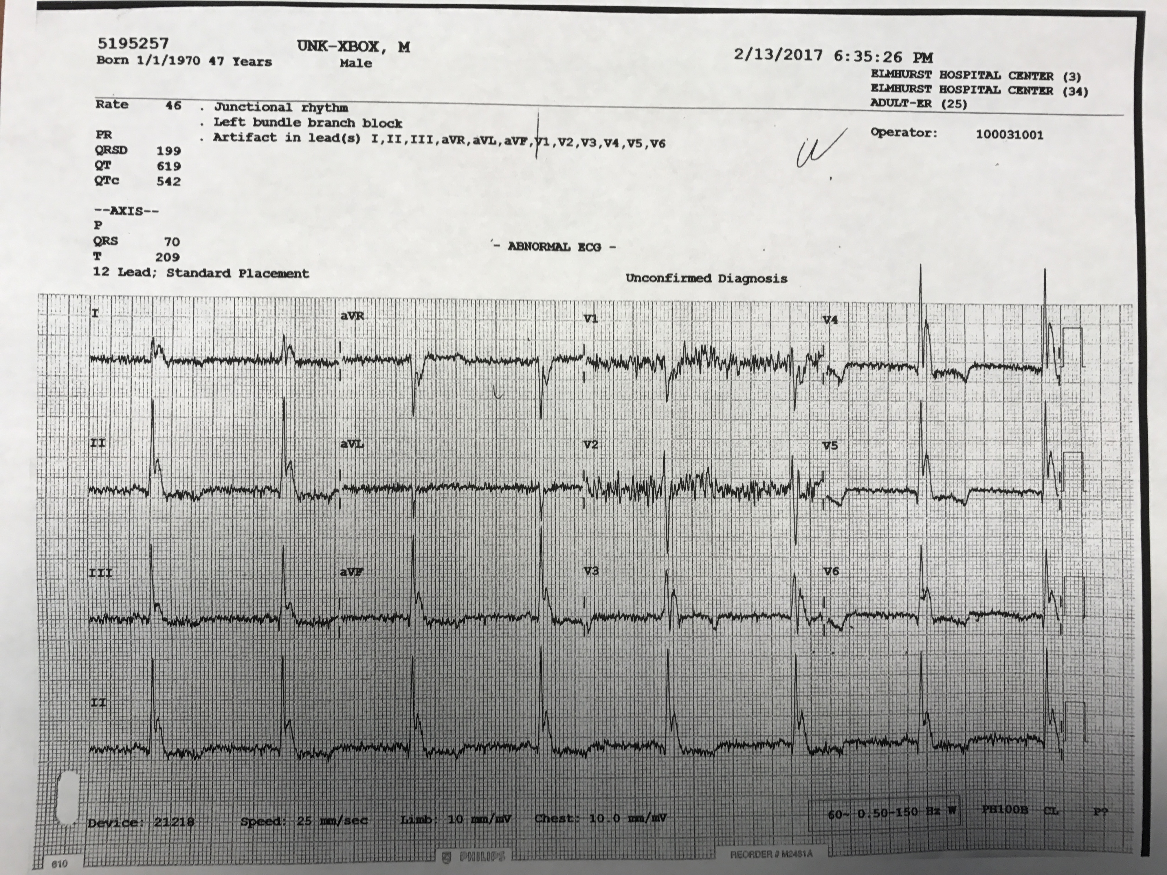
At first glance this this ECG demonstrates bradycardia which is likely sinus (though artifact makes identification of p-waves difficult), ST-depressions in leads V4-V6 and, most importantly, Osborn waves throughout. An Osborn wave, also known as a J wave, is an ECG finding most often associated with hypothermia. The Osborn wave has the appearance of a camel hump and is a positive deflection between the QRS complex and ST segments of the ECG. The magnitude of the Osborn wave usually correlates directly with the level of hypothermia and so should become less prominent as rewarming occurs. Other ECG findings that may be seen in hypothermia include bradycardia and prolonged PR, QRS and ST segments. When profound, hypothermia may also precipitate cardiac arrest. A reassuring ECG finding that is present here is the significant artifact seen throughout. This is secondary to shivering which is clearly desirable in the setting of profound hypothermia. As the patient’s core temperature increases you often see improvement or resolution of the bradycardia, Osborn waves, shivering artifact and prolongation of the different segments within the ECG. In an arresting patient, rewarming may be the trigger for return of circulation. When these ECG findings are present, it is absolutely essential that accurate temperatures be obtained. If hypothermia is identified decisive steps must be taken to rewarm the patient as this may be a lifesaving intervention.
Key Points:
The Osborn wave or J wave is a positive deflection between the QRS complex and ST segment of an ECG and has a camel hump appearance. It is most often associated with hypothermia.
The magnitude of the Osborn wave often correlates directly with level of hypothermia and so should improve as rewarming occurs.
Other ECG findings associated with hypothermia include bradycardia, wide PR, QRS and ST segments, and sometimes asystole or other cardiac arrest arrhythmias. These findings may improve or resolve with rewarming.
