Introduction:
The Focused Assessment with Sonography in Trauma (FAST) has been used by emergency physicians and surgeons for decades to diagnose hemoperitoneum and hemopericardium at the bedside. Many studies have demonstrated a good positive predictive value for the test in the setting of blunt torso trauma.
Focused Questions:
1. Is there fluid in the peritoneal cavity (ie. hemoperitoneum)?
2. Is there a pericardial effusion?
3. Is there fluid in the thorax (ie. hemothorax)?
4. Is there a pneumothorax? (see separate pneumothorax tutorial)
Required Views:
Video Overview:
Required Views:
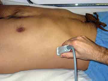
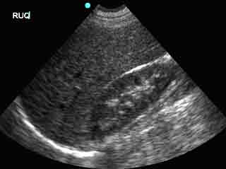
1. Right Upper Quadrant (Morrison’s Pouch)
| Probe position | Image |
 |
 |
| Notes | |
|
|
| Abnormal Studies | |
 
|
|
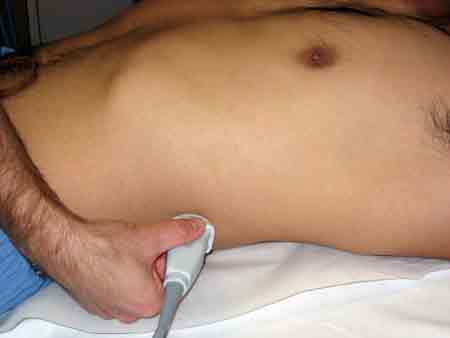
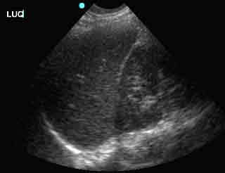
2. Left Upper Quadrant (Perisplenic space)
| Probe position | Image |
 |
 |
| Notes | |
|
|
| Abnormal Studies | |
 |
|
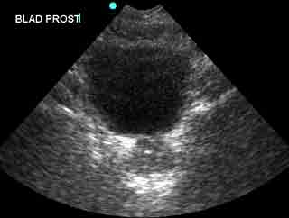
3. Rectovesicular space (Pelvic cul-de-sac)
| Probe position | Image |
 |
 |
| Notes | |
|
|
| Abnormal Studies | |
  |
|
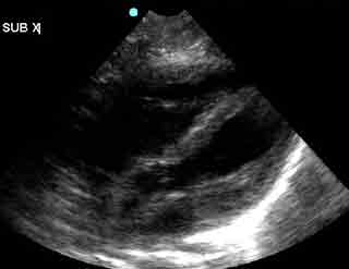
4. Pericardium
| Probe position | Image |
 |
 |
| Notes | |
|
|
| Abnormal Studies | |
 |
|


