Emergency physicians most frequently use pelvic ultrasound to confirm intrauterine pregnancy (IUP) in the setting of first trimester pregnancy complications. However estimation of gestational age is also described in ACEP’s ultrasound guidelines, and is worth discussing for a few reasons:
- Patients further along in their pregnancies are making their ways to Emergency Departments and it helps us guide pregnancy management (see I didn’t know I was pregnant)
- Emergency physicians are playing larger roles in Global Health/International Emergency care and are often among the first to volunteer in disaster situations.
- Accurate information on gestational age is helpful for the obstetricians who will follow up with these patients.
The type of measurement depends on the patient’s trimester with biometric data from the first trimester being the most accurate as there is very little biologic variation during this period.  Essentially, trust the dating from LMP if it is unequivocal and use ultrasonographic estimates if there is a discrepancy between the LMP & biometric estimates.
- 1st Trimester: Â >5 days
- 2nd Trimester: >7 days
Basics of Obstetric Scanning
- Ensure your ultrasound is on OB mode; this will provide access to all necessary calculations.
- As with all obstetric ultrasounds, first attempt a transabdominal approach, then if needed use transvaginal.
- All measurements should be taken at least three times and averaged.
- Estimations of gestational age in the third trimester are unreliable due to biologic variation with errors in estimates up to three weeks or greater.
Scanning: 1st Trimester
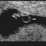
- Gestational Sac Measurement& Double Decidual sign:
- These findings represent the presence of hCG.
- Their presence is NOT definitive proof of an IUP.
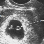
- Yolk sac:
- ~5th week (earliest embryonic structure)
- Fetal pole with fetal heart movement:
- ~6th week
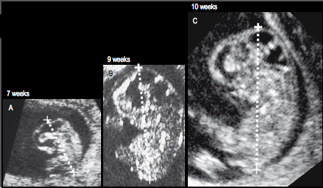
- Crown-Rump Length (CRL):
- Measurement can be performed after approximately 10 weeks gestation
- Fetus should be imaged in the longitudinal plane
- CRL is performed by placing the calipers at the the greatest embryonic length identified from the head to the rump.
- Do not include the yolk sac
- Accurate within 5 days.
Second & Third Trimester
- Biparietal Diameter (BPD) & Head circumference (HC). HC is the most accurate single measurement to help determine gestational age.
- Both are measuredthrough the same plane with the following landmarks:
- Anterior: frontal horns of the lateral ventricle, cavum septum pellucidum
- Central: thalami the third ventricle
- Posterior: occipital horns , cisterna venae magnae cerebri and insula
- BPD is measured with calipers placed at the outer margin of the proximal parietal bone to inner margin (to avoid overestimations due to posterior acoustic enhancement) of the distal parietal bone.
- HC is measured by placing the ellipse on the outside margin of the skull and expanding.
- Both are measuredthrough the same plane with the following landmarks:

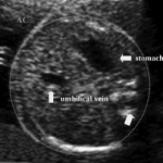
- Abdominal Circumference (AC)must be precisely measured at the following landmarks to avoid overestimations:
- Stomach and bifurcation of the main portal vein to into the right and left portal veins
- It should also be a perfectly round section
- AC measurement is made by placing the ellipse on the outside margin of the abdomen
- The smallest measurement obtained should be used
- This measurement has a high degree of intra and inter-observer variability
- Femur Length (FL):
- FL is measured from the origin (greater trochanter) of the distal end of the shaft (lateral condyle)
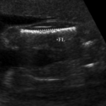
- The bone should be perpendicular to the beam
- Can be measured from 10 weeks onward.
- FL is measured from the origin (greater trochanter) of the distal end of the shaft (lateral condyle)
Pitfalls & Pearls:
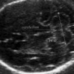
- This is not an exhaustive list of biometric measurements; others exist that are beyond the scope of this article, such as foot length, ear size, orbital diameter, cerebellum diameter etc.
- It is better to use an alternative measurement than use a poorly visualized structure.
- The first and earliest reliable measurements and estimates of gestational should be kept, and not changed on subsequent estimates. Â However if there are discrepancies between the gestational ages of two examinations, it may represent Intrauterine Growth Retardation (IUGR).
- In twin gestations, averages can be made between the measurements of twins
- Are EM physicians good at these measurements? Yes (see last two references)
References:
- Obstetric ultrasound : artistry in practice, Hobbins, John C. Blackwell Pub. 2008.
- Sonographic Determination of Gestational Age. Â Kalish RB, Chervenak F. Â TMJ. 2009; 59:2.202-208.
- Ultrasound in Obstetrics and Gynaecology (First Edition), Wladimiroff, Juriy. Eik-Nes, Sturla. Elsevier. 2009.
- Shah S et al. Accuracy of emergency physicians using ultrasound to determine gestational age in pregnant women. Am J Emerg Med. 2010 Sep;28(7):834-8. Epub 2010 Mar 26.
- Bailey C et al. Accuracy of emergency physicians using ultrasound measurement of crown-rump length to estimate gestational age in pregnant females. Am J Emerg Med. 2012 Feb 3.
