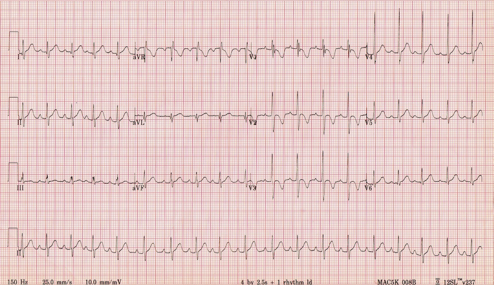
Take a look at the above EKG. If you saw this in a 70 year old patient with chest pain, you might be a bit concerned. Tachycardic, inverted T waves anteriorly, a partial RBBB in V1. Are these ischemic changes going on? Where is this man’s prior EKG?! But wait, here’s the good news. The above EKG actually belongs to a 2 year old patient. Why did we even get an EKG? Can 2 year olds even say chest pain?
Now, the pediatric EKG can be challenging because we don’t see them as often and normal just looks different than what we are used to as ED doctors. Kids are not little adults, and baby hearts are definitely not…little adult hearts.
The different anatomy and physiology of infants account for many of the different findings on EKG. At birth, the right ventricle is thicker and larger than the left ventricle due to the fetal circulation system in utero. This results in an EKG that looks like R ventricular hypertrophy in the adult, including a rightward axis and flipped T waves in V1-3. Many of these changes will be replaced by adult EKG findings by adolescence, but until then, we need to be familiar with some of the findings on a pediatric EKG.
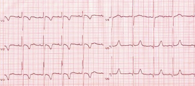
Juvenile T wave pattern refers to the T wave inversions commonly seen in the anterior leads. In the first week of life, the T waves are all upright. In the 2nd week, the T waves in V1-3 invert and will remain that way until adolescence when the heart begins to assume a more familiar configuration as an adult. Persistent juvenile T wave pattern is worth mentioning as a possible normal variant in young adults. This refers to T wave inversions similar to juvenile pattern that persists into adulthood. It is most commonly associated with African American women up to the age of 30. The T waves are asymmetric, shallow (<3mm), and limited solely to V1-3 (see below EKG).
Now, once we start talking about adult EKGs, flipped T waves are a whole other story. They can be normal variants, such as in persistent juvenile t wave pattern. But, in the appropriate context, keep an eye out for the usual dangerous suspects such as ACS, PE, and even Wellens syndrome.
Thanks to Dr. Jenny Sanders who gave a great talk on pediatric EKGs yesterday at conference. You can find the full lecture on our SinaiEM Vimeo page and soon to be uploaded onto our website at www.SinaiEM.org.
Sources
http://lifeinthefastlane.com/ecg-library/paediatric-ecg-interpretation/
http://lifeinthefastlane.com/ecg-library/basics/t-wave/
http://hqmeded-ecg.blogspot.com/2015/01/persistent-juvenile-t-wave-pattern.html
