Introduction:
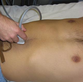
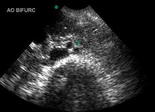
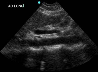
Many studies have demonstrated the accuracy of bedside ultrasound in detecting abdominal aortic aneurysm. Since the majority of these aneurysms occur distal to the renal arteries, it is imperative that the entire abdominal aorta (down to the aortic bifurcation) is visualized. Measurements should be taken across the widest diameter of aorta, from outer wall to outer wall.
Focused Questions:
- Is the aorta less than 3cm?
- Are the iliac arteries less than 1.5cm?
Video Overview:
Required Views:
1. Proximal Aorta (transverse)
| Probe position | Image |
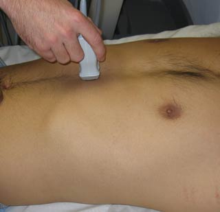 |
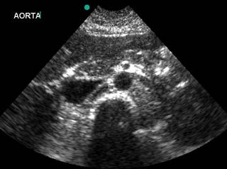 |
| Notes | |
|
|
| Abnormal Studies | |
 |
|
2. Mid Aorta (transverse)
| Probe position | Image |
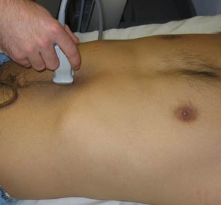 |
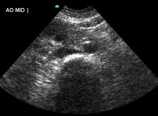 |
| Notes | |
|
|
| Abnormal Studies | |
 |
|
3. Aortic bifurcation/iliac arteries (transverse)
| Probe position | Image |
 |
 |
| Notes | |
|
|
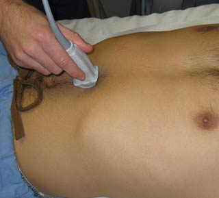
4. Mid Aorta (longitudinal)
| Probe position | Image |
 |
 |
| Notes | |
|
|
| Abnormal Studies | |
 |
|
