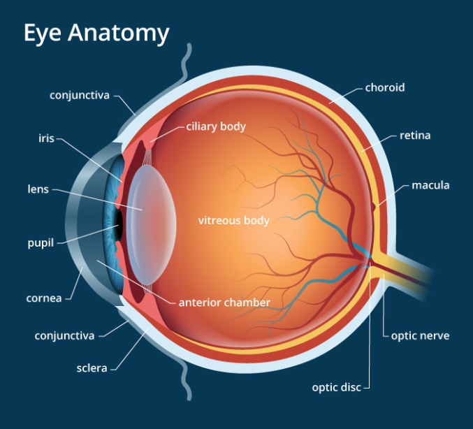
THE EYE EXAM

- Keep it basic…
- APD
- Intra-ocular Pressures: Tono-pen v Applanator (Goldmann)
- Visual Acuity
- or be a Slit Lamp KWEEN
- Move outside in: Lids → Eyeball
- Lids: ducts, eyelashes, orbital lesions or findings
- EYE: Full EOM assessment and conjunctival assessment
- Anterior:
- Cornea for opacity, irregularities, fluericin staining for abrasions/ulcerations.
- Anterior chamber assessment for “cell and flare” or hypopyon.
- Lens for opacities.
- Assess for extrusion of IOC contents.
- Posterior: the dilated exam. Save that for your optho friends — but Ocular US for optic nerve measurements is a neat trick that we can do.
- Visualize the posterior eye indirectly to assess for retinal detachment vs vitreous hemorrhage
- Visualize & measure the optic nerve (3mm deep, with 5mm as your upper limit of normal)
- Anterior:
- Applanation for pressure (Goldmann is the GOLD standard…hehehe)
- Here’s a quick readable how-to: https://www.ncbi.nlm.nih.gov/pmc/articles/PMC2206330/
- And these are youtube demonstrations:
- https://www.youtube.com/watch?v=mS2HvAN4Uzg
- https://www.youtube.com/watch?v=0b2Mv54mQcs (start at 07:48)
